- About the Lab
- Member
- Equipment
- Protocol
 The Histopathology & Bio-imaging Lab provides techniques, equipment and protocols for bio-imaging and non-destructive 3-D imaging evaluation as well as histopathological examination and histomorphometric analysis for musculoskeletal research and for developing innovative drugs in bone and joint diseases. So far, the lab has established the following techniques.
Ø Biophotonic-based Fluorescence Imaging Techniques
Lumina XR Series III could sensitively image bioluminescent / fluorescent reporters and differentially evaluate their distribution in various organs within the same animal. It is employed to examine drug delivery by in vivo bio-imaging analysis.
Ø X-ray-based Three-dimensional Imaging Techniques
Scanco VivaCT 40 could provide non-destructive imaging and analysis of musculoskeletal tissue. It is employed to perform in vivo and ex vivo measurement of bone and cartilage specimens for micro-architecture.
Ø Histopathological Examination & Histomorphometric Analysis & Gene Expression Analysis in
specific cells
Non-decalcified histological processing for bone tissue and static / dynamic bone histomorphometric analysis could be performed in the lab. Techniques of wide special stains for bone and cartilage, such as the VonKossa, tartrate-resistant acid phosphatase (TRAP), alkaline phosphatase, Gömöritrichrome, safranin O and toluidine blue stains are provided in the lab. Further, laser-captured micro-dissection in combination with Q-PCR analysis for gene expression in specific cells is also established in the lab.
Ø Planar Radiographic Imaging Techniques
The Faxitron MX-20 radiography system can be used for a broad range of bone studies including: bone metastasis, osteoporosis, phenotyping, and bone growth / regeneration studies.
◆ Research Assistant Professor
◆ Major: Clinical Orthopedics & Musculoskeletal Science
◆ E-mail: borisguo@hkbu.edu.hk

◆ Ph.D. Student
◆ Major: Orthopedics Surgery
◆ E-mail: drlausums@gmail.com

Ø Equipment Name: EXAKT® Cut / Grinding System
Ø Location: Rm. 609, SCM
Ø Technician: Dr. Baosheng Guo (borisguo@hkbu.edu.hk)
Ø Description: The EXAKT Cut / grinding System (EXAKT Technologies, Inc. Germany) is composed of five individual
devices (E300C, E400, E401 / 40, E510 and E520), which work inconcert to produce thin sections of bone / joint
samples.
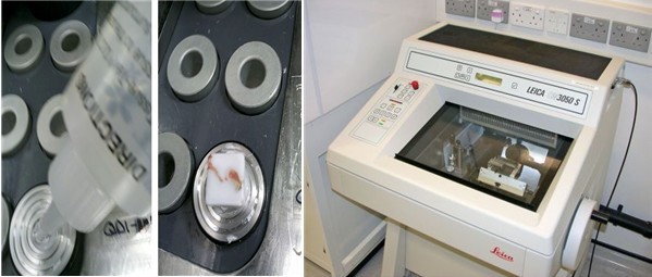
Ø Equipment Name: Leica Cryostat
Ø Location: Rm. 702, SCM
Ø Technician: Mr. Michael Wong (whwong@hkbu.edu.hk)
Ø Description: The Leica Cryostat facilitates preparing frozen sections from decalcified bone tissue for observation under
a microscope.
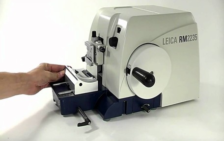
Ø Equipment Name: Leica Rotary Microtome
Ø Location: Rm. 607, SCM
Ø Technician: Mr. Michael Wong (whwong@hkbu.edu.hk)
Ø Description: Leica Rotary Microtome were used for routine sectioning of paraffin-embedded tissues for observation
under a microscope.
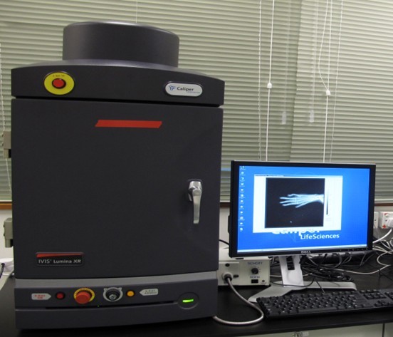
Ø Equipment Name: The Lumina XR Series III
Ø Location: Rm. 602, SCM
Ø Technician: Ms. Nickie Chan (nickie@hkbu.edu.hk)
Ø Description: The Lumina XR Series III pre-clinical in vivo imaging system offers precise optical and X-ray overlay to
bring the optical signal into anatomical context. Perkin Elmer Life Science’s Living Image® software automates all
the controls, settings and analysis.
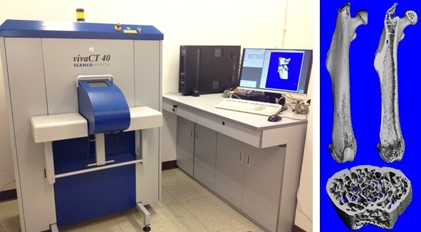
Ø Equipment Name: SCANO® vivaCT 40
Ø Location: Rm. 601, SCM
Ø Technician: Mr. Michael Wong (whwong@hkbu.edu.hk)
Ø Description: VivaCT 40 is a fast and flexible in vivo microCT for preclinical studies. The low X-ray dose allows serial
measurements of individuals. Whole-body scans or scans from selected regions of mice can be made, while head
and hind limbs of larger rodents can be scanned using beds supplied with the system.

Ø Equipment Name: BIOQUANTOSTEO Analysis System
Ø Location: Rm. 601, SCM
Ø Technician: Dr. Baosheng Guo (borisguo@hkbu.edu.hk)
Ø Description: The BIOQUANT OSTEO software enables cutting-edge bone biology research in animal models and
human biopsy. It supports both high-throughput automation and precise manual interaction for ASBMR standard bone
histomorphometry in digital histology and microCT. Unique tools simplify skeletal phenotyping, characterize arthritis
models, help quantify cancer metastasis, measure chondrocyte proliferation aid in forensic anthropology and
quantify implant osseointegration.

Ø Equipment Name: Laser Captured Microdissection
Ø Location: Rm. 609, SCM
Ø Technician: Mr. Michael Wong (whwong@hkbu.edu.hk)
Ø Description: Laser Capture Microdissection system (LCM, PixCell IIe from Aucturus) provides one of the best tools
available to date for isolation of individual cells or specific populations of cells, or bacteria of interest from tissue
sections for further molecular and biochemical analysis.
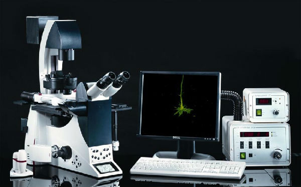
Ø Equipment Name: Fluorescent microscope with image acquisition system
Ø Location: Rm. 602, SCM
Ø Technician: Mr. Michael Wong (whwong@hkbu.edu.hk)
Ø Description: The Leica AF6000 advanced fluorescence imaging systems are ideal for applications in fluorescence
microscopy and image analysis including live cell time-lapse experiments, multi-positioning, z-stacking and
deconvolution. The versatile, fully integrated solutions offer harmony between hardware and software and combine
the ultimate speed, reliability, and facilities for experiment design, analysis and operational convenience.

Ø Equipment Name: Axioplan 2 and Axiophot 2 Universal Microscopes
Ø Location: Rm. 604, SCM
Ø Technician: Mr. Michael Wong (whwong@hkbu.edu.hk)
Ø Description: Zeiss axioplan 2 automated / digital imaging microscope system w/tmc vibration isolation table: zeiss
fluar 20x, zeiss fluar10x, zeiss fluar 2.5x, zeiss plan neofluar 1.25x, pl 10x / 25 lenses,zeiss axio camera, ebq 100
isolated illuminator, lep mac 5000 ps automation controller system (ludl eletronic products, inc.), extended travel lep
stage for loading mulitple slides, tmc isolation table.
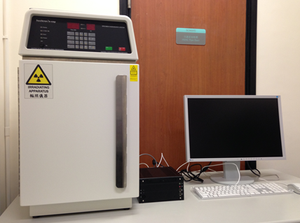
Ø Equipment Name: Faxitron MX-20 Digital Radiography System
Ø Location: Rm. 601, SCM
Ø Technician: Dr. Baosheng Guo (whwong@hkbu.edu.hk)
Ø Description: The cabinet X-ray system provides high-resolution planar x-ray images of mice and rats quickly and
affordably. The energy of the X-ray tube ranges from 10-35KV with a maximum tube current of 300uA. The focal
spot is 20 micrometers allowing for very high spatial resolution.
 Protocol for fluoresce double labeling

